 My biopark
WELCOME TO BIOPARK
What's in
the news ?
RUE ClÉMENT ADER, 10 6041 CHARLEROI
+32 71 91 99 50


News
The nucleolus: a strong biomarker of cancer
February 27th 2020
Candice Leblanc
The ULB spin-off project RIBOcancer, funded by the FIRST Spin-off programme of the Walloon Region, is developing a new cancer diagnostic and prognostic method based on the quantitative analysis of nucleoli, which are valuable indicators of cell health.
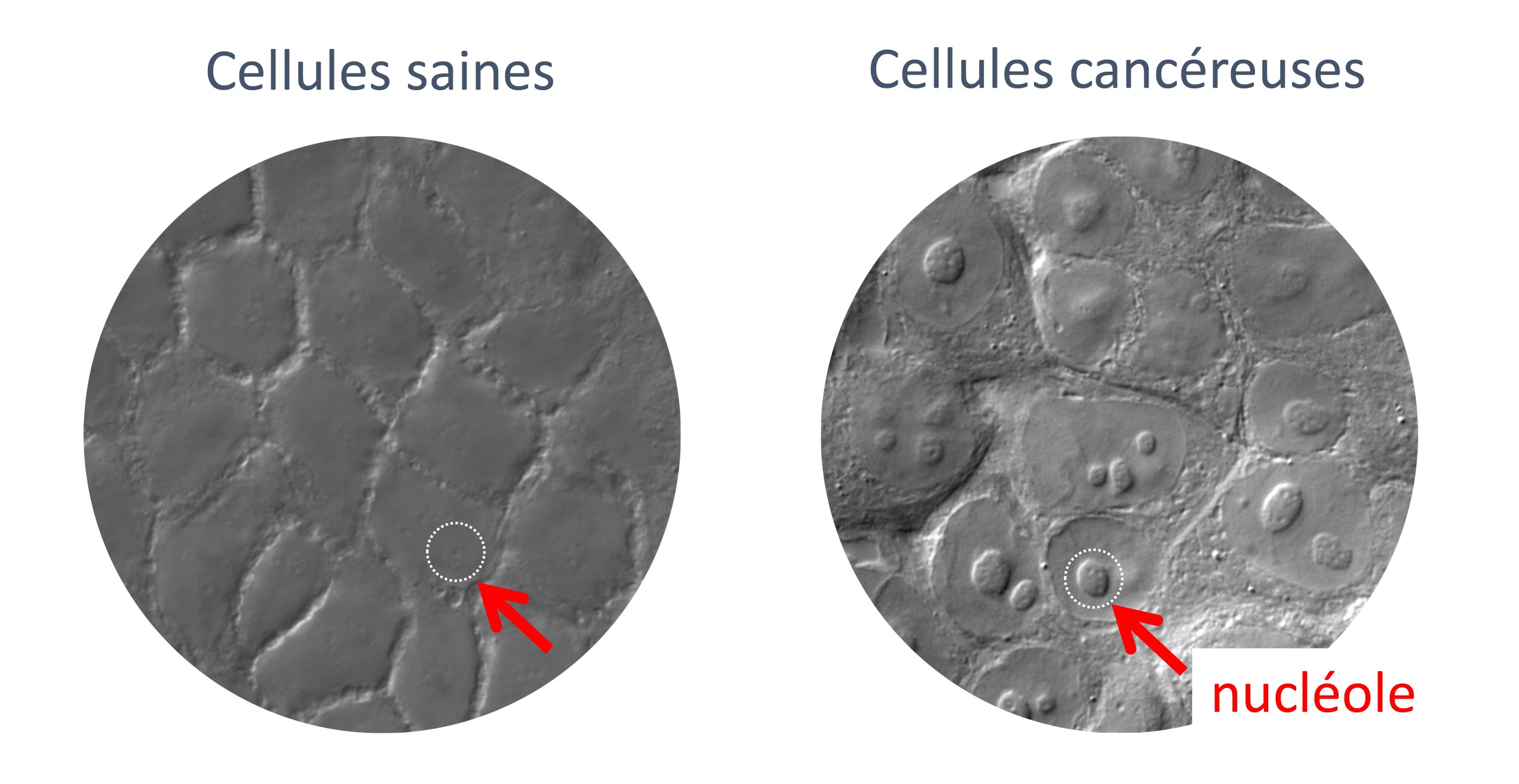
Fig. 1: The size, shape and number of nucleoli (i.e. the ribosome ‘factories’) in each cell are indicators of the health status of a cell. The illustration shows a comparison between healthy and cancerous cells.
Inside the nucleus of each cell are nucleoli. These small ‘factories’ are sites of ribosomes production and assembly; in turn, the ribosomes build proteins. ‘By looking at the size, shape and number of nucleoli, we can determine whether a cell is healthy or not’, explains Professor Denis Lafontaine, research director at F.R.S./FNRS, professor at ULB and head of the RNA Molecular Biology Laboratory. ‘In a cancerous cell, for instance, nucleoli are generally larger, more numerous and deformed. This makes nucleolar morphology an excellent biomarker.’
Using an algorithm to ‘score’ nucleoli
Nucleoli can easily be seen in a microscope, and have been known to scientists for a long time. However, with no appropriate quantitative tools available, their use as biomarkers in clinical anatomopathology has remained limited. But this could change soon! ‘We have recently developed a detection method and an algorithm that analyses the morphology of nucleoli with very high accuracy and, more importantly, calculates a “score of nucleolar disruption” (1)’, explains Professor Lafontaine. ‘This method, called the iNo score, has been successfully tested on cell models. We are currently working on a proof of concept using human biopsies.’
Detecting anomalies at an earlier stage
The iNo score could be an improvement over existing diagnostic approaches, for instance in the case of kidney cancers. ‘The grade of kidney cancers is already determined by the size of the nucleolus, but our method is more accurate. Our software not only measures the size of the nucleolus, it also detects structural abnormalities that are too limited for pathologists to see “with the naked eye”. Current morphological biomarkers are often based on cells that have already been deformed by cancer. Our software can spot abnormalities earlier, at the intracellular level, when the overall cell morphology is still intact.’ This means that the iNo scoring method may be able to detect cancer at an earlier stage.
Applications and prospects
RIBOcancer currently receives support from the Walloon Region for the translational development of the iNo scoring method. This implies making it compatible with clinical laboratories and use on human tissues. ‘In the next few months, we must raise significant funds in order to continue building our team, finalize the technological developments , and explore all the potential business opportunities offered by our method’, concludes professor Lafontaine. ‘We are looking into a number of possibilities: including a “ready-to-use” diagnostic kit, sell licenses to use the iNo scoring software, create a reference clinical laboratory or a cloud service for pathologists, and so on.’ Investors wanted!
Notes:
- This discovery has been the subject of two articles in the scientific journal Nature, as well as of one patent.